Description
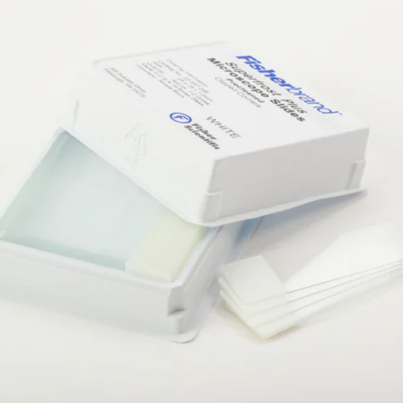
Superfrost® Plus Microscope Slide
The Superfrost® Plus Microscope Slide is a premium microscopic slide designed for reliable preparation, handling, and long-term retention of tissue and cytology specimens in clinical and research laboratories. Engineered for compatibility with routine histology workflows, the slide features a positively charged surface that promotes strong adhesion of formalin-fixed, paraffin-embedded (FFPE) tissue sections, frozen sections, and many cytology preparations. This enhanced adherence helps reduce section loss during demanding staining and processing steps, supporting consistent results across routine and advanced applications.
Purpose and applications
Superfrost® Plus slides are intended for mounting and staining thin specimen sections for light microscopy. The positively charged surface is particularly useful for procedures that include multiple aqueous steps, heat, antigen retrieval, or enzymatic treatment, where standard slides may show increased risk of tissue lift. These slides are commonly used in hematoxylin and eosin (H&E) staining, special stains, immunohistochemistry (IHC), and in situ hybridization (ISH) methods when strong specimen adhesion is desired.
Key features
- Positively charged surface to support firm specimen attachment and minimize tissue detachment during staining and retrieval steps.
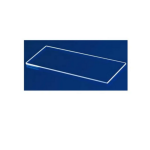
- Consistent optical quality suitable for routine brightfield microscopy and high-resolution evaluation.
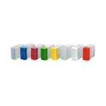
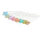
- Frosted labeling end to enable clear specimen identification using pencils or laboratory markers and to support organized slide management.
- Clean, ready-to-use format appropriate for standardized laboratory workflows.
- Broad workflow compatibility with common microtomy, cryostat, staining, and coverslipping procedures.
Laboratory performance benefits
By improving section adherence, Superfrost® Plus slides can help laboratories maintain slide integrity through repetitive wash cycles and high-temperature or high-pH antigen retrieval protocols. This can reduce the need for repeat cutting and restaining, preserve limited patient material, and support more efficient turnaround times. Strong adhesion also aids in maintaining section morphology and positional stability, helping technologists and pathologists interpret staining patterns with confidence.
Handling and use considerations
For best results, handle slides by the edges or frosted end to reduce contamination and fingerprints on the viewing area. Ensure sections are placed flat and free of folds, then dry according to laboratory protocol before proceeding with staining. Adhesion performance may vary with specimen type, fixative, section thickness, and processing conditions; laboratories should validate slides for their intended methods, including any IHC or ISH protocols and antigen retrieval conditions.
Typical specifications
| Attribute | Description |
|---|---|
| Product type | Microscope slide for histology and cytology |
| Surface | Positively charged (enhanced specimen adhesion) |
| Labeling area | Frosted end for identification |
| Use | Mounting and staining of specimen sections for microscopic examination |
Summary
The Superfrost® Plus Microscope Slide is a dependable choice for laboratories seeking improved tissue retention and robust slide performance across routine histology and advanced staining procedures. Its positively charged surface and practical labeling area support consistent specimen preparation, efficient workflow, and high-quality microscopic evaluation.