Description
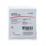
Colorfrost® Plus Microscope Slide
Colorfrost® Plus Microscope Slides are premium microscopic slides designed for reliable preparation, staining, and long-term preservation of tissue and cytology specimens in clinical, research, and teaching laboratories. Built for consistent performance across routine histology and specialized staining workflows, these slides support clear specimen visualization under brightfield and related microscopy techniques while helping streamline identification and handling.
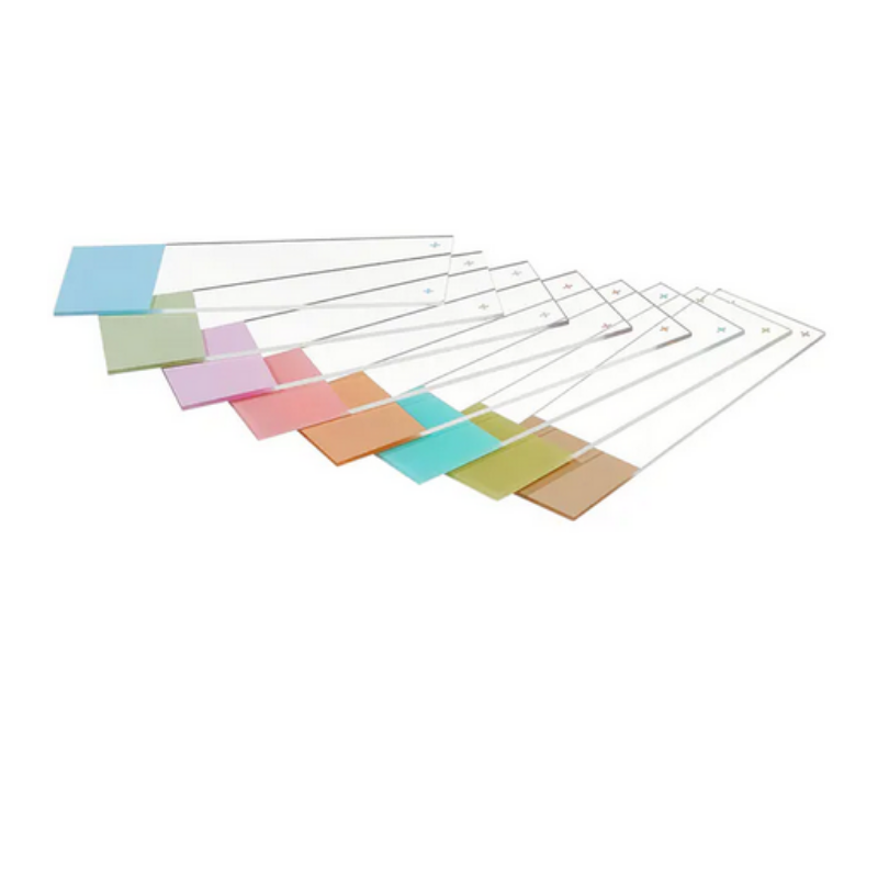
Each slide is manufactured from high-quality glass to provide excellent optical clarity, a flat viewing surface, and uniform thickness that supports consistent focusing and image capture. Polished edges and carefully finished corners promote safer handling, reduce the likelihood of glove tears, and help protect microtome sections and coverslips during processing and transport.
The defining feature of Colorfrost® Plus is an enhanced, positively charged specimen-adhesive surface that improves section retention during demanding procedures. This adhesion helps minimize tissue lift, section loss, and edge curling during heat exposure, antigen retrieval, enzymatic digestion, and multiple wash steps commonly encountered in immunohistochemistry (IHC), in situ hybridization (ISH), and special stains. The result is improved workflow efficiency, reduced repeat cutting, and more dependable slide-to-slide results.
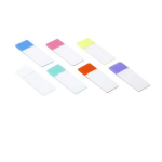
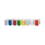
A colored, frosted end provides a dedicated labeling area that supports clear sample identification and orientation. The frosted writing surface accepts common laboratory marking methods and is designed to remain legible through routine handling. Color coding can aid organization by case, stain type, or priority, helping reduce mix-ups and improving traceability in multi-step processing environments.
Key Features
- Positively charged adhesion to promote strong tissue and cell attachment through rigorous staining protocols.
- High optical clarity for sharp viewing and consistent imaging.
- Uniform dimensions and thickness to support reproducible microscopy and automated slide handling.
- Colored frosted label area to improve readability and workflow organization.
- Polished edges for safer handling and reduced risk of specimen disruption.
Typical Applications
- Routine histology (H&E) and cytology preparations
- Immunohistochemistry (IHC) and immunocytochemistry (ICC)
- In situ hybridization (ISH) and related molecular pathology workflows
- Special stains requiring multiple incubations and washes
- Research microscopy, teaching sets, and archivable slide collections
Workflow Benefits
By improving specimen adherence, Colorfrost® Plus Slides help protect valuable samples, reduce rework, and support more consistent staining quality. The dependable label area helps maintain specimen identity from sectioning through coverslipping and storage, supporting good laboratory practice and documentation requirements.
Handling and Storage
- Handle slides by the edges to maintain a clean viewing area.
- Label within the frosted region before or after section placement per laboratory protocol.
- Store slides in a clean, dry environment and use appropriate slide holders to protect surfaces.
- Follow validated laboratory procedures for fixation, baking, staining, and coverslipping.
Colorfrost® Plus Microscope Slides are suited for laboratories that require dependable adhesion, clear labeling, and consistent optical performance for high-quality microscopic evaluation.